Grafik
Plate X
In this Plate we have a full section of the Brain. Showing chiefly the great relations of the parts, the relative places of the Ventricles, and their communication, the Arteries of the Corpus Callosum, and the Falx and Sinuses. To make those parts sufficiently minute, it has been necessary to draw them of the full size. a. a. a. The Cranium, cut perpendicularly a little to the left of the great longitudinal Sinus. b. b. The Falx formed by the Dura Mater, and descending betwixt the hemispheres of the Cerebrum, reaching anteriorly from the Jr' Crista Gali of the Sphenoid Bone backwards, and deepening as it runs back, until it is infixed or continued into the Tentorium, by which both these partitions are kept true, and mutually depending on each other. c. A Thread holding out the cut edge of the Tentorium Cerebelli Super Extensum. D. The cut Edge of the Tentorium, which stretches nearly horizontally over the Cerebellum, and supports the posterior Lobe of the Cerebrum. E. The Surface of the right hemisphere of the Cerebrum, as it appears under the Falx, that partition not descending quite to the Corpus Callosum. F. The Section of the Corpus Callosum. g. The Lower Surface of the Corpus Callosum. H. The Septum, dividing the left lateral Ventricle (which is here laid open, and is of course under shadow) from the right lateral Ventricle. I. The Upper Surface of the Fornix, where it is forming the posterior Crus. K. The inferior Surface of the Fornix, that which is called Lyra. L. The anterior left Crus of the Fornix, under which is the opening of the right lateral Ventricle, which of course forms a communication with the left lateral, and the third Ventricle, at the same time. M. The anterior Comissure of the Brain, which is truly a medullary body, running transversely and connecting the Hemispheres. N. The Prominence made by the termination of the right anterior Crus of the Fornix. O. The Opening of the right lateral Ventricle into what is described by many Authors, as the most anterior part of the third Ventricle ; by others, as the Foramen Commune Anterius ; which indeed conveys the most accurate idea of this part, for it is a space under the Anterior Crura of the Fornix, into which both the lateral Ventricles open, and which therefore makes a communication betwixt them. By others, it is called Vulva, from its appearance upon raising the Fornix in the usual manner of dissecting the Brain, p. The third Ventricle. The remains of the Comissura Mollis are scarcely to be observed after the separation of the Thalami Nervorum Opticorum—therefore it is not represented in the draw ing ; but we can understand that it is the union of the Thalami above the letter P. and that the space under it is the third Ventricle. This is a gutter-like cavity communicating or continued into that common space under the anterior Crura of the Fornix, and at the same time opening downwards into the Infundibulum, and backwards by the Iter ad quartum Ventriculum. q. A Probe, introduced from the bottom and fore part of the third Ventricle into the Infundibulum, and which is here represented as reaching nearly to the surface of the Glandula Pituitaria. r. The Glandula Pituitara, seated in the Sella Turcica, s. The Iter ad quartum Ventriculum. t. The Comissura Posterior, the connection of which with the Pineal Gland is accurately represented, v. The Pedunculi of the Pineal Gland prolonged upon the Thalamus Nervi Optici. u. The Tubercula Quadrigemina, or Nates and Testes, w. Valvula Vieussenii. x. The Pineal Gland, y. The Cavitiy of the Fourth Ventricle. z. The Calamus Scriptorius. a. a. The Cerebellum in outline, lying deep in the Scull-cap, and under the Tentorium. b. The Tuber Annulare, or Pons Varolii. c. The Medulla Oblongata, both of these in outline. d. The Pia Mater, closing up the lower part of the fourth Ventricle. ARTERIES, VEINS, AND SINUSES. 1. The Internal Carotid Artery, passing into the Cranium through the Sphenoid bone. 2. The Internal Carotid within the Scull. 3. The Anterior Cerebral Artery of the right side. 4. The left Anterior Artery of the Cerebrum. 5. The Continuation of the Right Cerebral Artery on the other side of the Falx, chiefly. 6. The continued Trunk of the left Cerebral Artery. 7. The Artery of the Corpus Callosum, sent off from the last mentioned Artery, which running along the arch of the Corpus Callosum, is distributed to the loose texture of the Pia Mater. 8. 8. 8. 8. The Longitudinal Sinus seen in its whole length. 9. 9. 9. The superficial Veins of the Cerebrum, entering the longitudinal Sinus. 10. The Lateral Sinus of the left side, where it is opened by the general section.—Here then is a union of the great Sinuses of the Brain. 11. The Fourth Sinus, which lies betwixt the angle of the union of the Falx and Tentorium. 12. The Fifth Sinus, or Inferior Longitudinal Sinus, running upon the edge of the Falx, or (taking the similitude from which the word Falx is borrowed) upon the cutting edge of the sickle. 13. The Commencement of this Inferior Longitudinal Sinus, by small veins arising from the Corpus Callosum, where it forms some beautiful inosculations. 14. At this place the Inferior Longitudinal Sinus, which can be scarcely considered in any other light than as a vein anterior to this, enters the firm investure of the Dura Mater, forming the Falx. 15. 16. 17. Veins running beautifully tortuous in the Falx, and forming frequent communications with the superior and inferior Longitudinal Sinuses.
- Standort
-
Universitätsbibliothek Heidelberg
- Sammlung
-
UB Anatomische Illustrationen
- Inventarnummer
-
P 1242-4 Folio RES
- Sprache
-
Englisch
- Klassifikation
-
Stich (Gattung)
- Bezug (was)
-
Anatomie
Gehirn
Illustration
- Bezug (wer)
- Ereignis
-
Herstellung
- (wer)
- (wo)
-
London
- (wann)
-
um 1802
- Ereignis
-
Veröffentlichung
- (wer)
-
Longman
- (wann)
-
1802
- Ereignis
-
Veröffentlichung
- (wo)
-
London
- Letzte Aktualisierung
-
26.03.2025, 09:43 MEZ
Datenpartner
Ruprecht-Karls-Universität Heidelberg. Universitätsbibliothek. Bei Fragen zum Objekt wenden Sie sich bitte an den Datenpartner.
Objekttyp
- Grafik
Beteiligte
Entstanden
- um 1802
- 1802
Ähnliche Objekte (12)
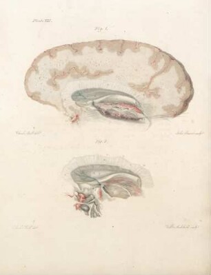
Plate VIII
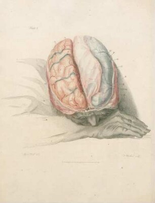
Plate I
Plate VI
Plate IX
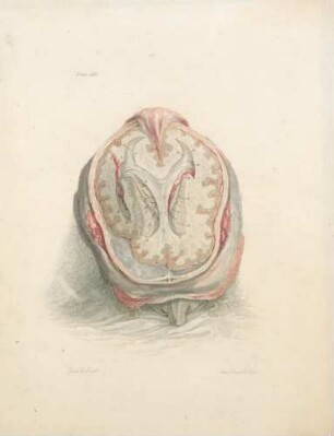
Plate III
Plate V
Plate IV
Plate XI
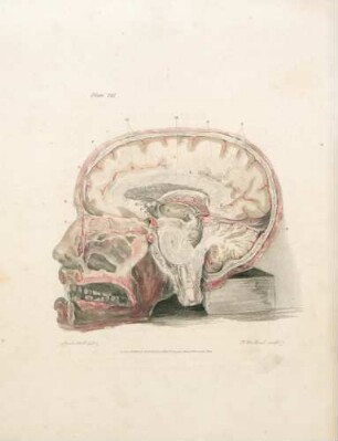
Plate VII
Plate II
Plate XII

21: The quarterly journal of pure and applied mathematics
Plate VIII
Plate I
Plate VI
Plate IX
Plate III
Plate V
Plate IV
Plate XI
Plate VII
Plate II
Plate XII
21: The quarterly journal of pure and applied mathematics
Plate VIII
Plate I
Plate VI
Plate IX
Plate III
Plate V
Plate IV
Plate XI
Plate VII
Plate II
Plate XII