Grafik
Plate V
In this Plate the Brain alone is represented. It shows the Choroid Plexus, and Velum Interpositum of Haller raised, the Fornix being'taken away, and the Corpora Striata; the Centrum Semicirculare Geminum; the Thalami Nervorum Opticorum; and the Pineal Gland. a. The Corpus Striatum: The Brain being now out so far down as to show the intermingling of the cineritious and medullary matter. b. b. The Thalami Nervorum Opticorum, which were almost intirely covered by the Fornix and Choroid Plexus. c. TIicTenia Semicircularis, or Tenia Striata. See Note. d. The Comissura Mollis, or union betwixt the Thalami Nervorum Opticorum, which leaves upon the fore and back part an opening into the third Ventricle. e. e. The Posterior Sinus of the lateral Ventricle. f. The Hippocampus Minor. g. The Cornu Ammonis, or Hippocampus. h. The Velum Interpositum, or Toile Choroidienne, held up by a ligature at the point of union betwixt the two plexus of the lateral Ventricle, and shewing the manner in which they are continued into a delicate plexus, which runs backwards upon the lower surface of the Velum, and which may be observed to split again, and involve the Pineal Gland. i. The Pineal Gland; Connected with the Velum, surrounded by the branches of the veins, and pulled from its seat by the lifting of the Velum. The Velum must be completely raised, and held back before the Pineal Gland can be seen in this view of the parts. k. The Foramen Commune Anterius, or Vulva. l. The Anus. m. The Anterior Comissure of the Cerebrum. Upon separating gently the Thalami Nervorum Opticorum, we see in a fresh subject the cohesion formed by the Comissura Mollis. It is from not having observed this union that authors have described this as the third Ventricle. In most of our difficulties let us return to Vieusscns, and we shall find, in a few words, the simple truth. It is a rima, or gutter-like cavity, under the Comissura Mollis, upon the anterior part of which, and under the Vulva, we see the beginning of the Infundibulum, and on the back part the Iter ad quartum Ventriculum.
- Location
-
Universitätsbibliothek Heidelberg
- Collection
-
UB Anatomische Illustrationen
- Inventory number
-
P 1242-4 Folio RES
- Language
-
Englisch
- Classification
-
Stich (Gattung)
- Subject (what)
-
Anatomie
Gehirn
Illustration
- Subject (who)
- Event
-
Herstellung
- (who)
- (where)
-
London
- (when)
-
um 1802
- Event
-
Veröffentlichung
- (who)
-
Longman
- (when)
-
1802
- Event
-
Veröffentlichung
- (where)
-
London
- Last update
- 15.09.2030, 10:43 AM CEST
Data provider
Ruprecht-Karls-Universität Heidelberg. Universitätsbibliothek. If you have any questions about the object, please contact the data provider.
Object type
- Grafik
Associated
Time of origin
- um 1802
- 1802
Other Objects (12)
Plate VIII
Plate X
Plate I
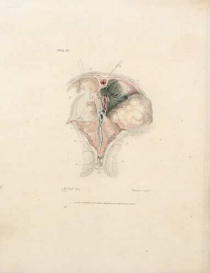
Plate VI
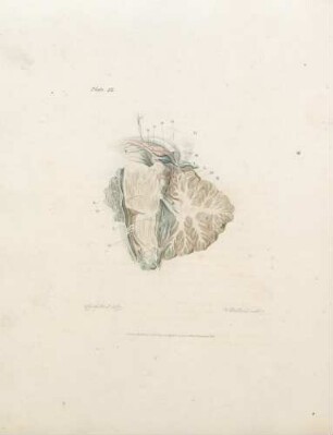
Plate IX
Plate III
Plate XII
Plate IV
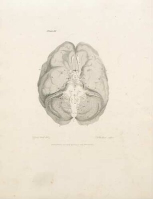
Plate XI
Plate VII
Plate II

21: The quarterly journal of pure and applied mathematics
Plate VIII
Plate X
Plate I
Plate VI
Plate IX
Plate III
Plate XII
Plate IV
Plate XI
Plate VII
Plate II
21: The quarterly journal of pure and applied mathematics
Plate VIII
Plate X
Plate I
Plate VI
Plate IX
Plate III
Plate XII
Plate IV
Plate XI
Plate VII
Plate II