Grafik
Plate XII
This Plate shews the Base of the Cranium, the place of the great Arteries and Sinuses, and the exit of the Nerves from the Scull. By comparing it with the last we shall learn the relation of the base of the Encephalon to the base of the Scull. a. The Frontal Sinuses. b. b. The Cranium. c. c. The most elevated part of the base of the Scull, formed by the Orbital Plate of the Frontal Bones, upon which the Anterior Lobes of the Brain, Plate XI. a. a. rest. d. The Crista Galli of the ./Ethmoid Bone, upon which the anterior part of the Falx, Plate XI. b. b. takes firm origin. e. The dura Mater turned back a little from its adhesion to the Frontal Bone. f. f. The acute edge of that part of the Sphenoid bone called the Wing of Ingrassias, which enters into the Fossa Silvii. (Plate XI. n. n.) g. g. The Fossa formed by the Temporal and Sphenoid Bones for lodging the Middle Lobe of the Cerebrum. Plate XI. b. h. The Tentorium upon which the Posterior Lobe of the Cerebrum, Plate XI. c. rests, I. i. The deep hollow formed by the Occipital Bone for the lodgment of the Cerebellum, Plate XI. i. d. k. ARTERIES. k. k. The Internal Carotid Artery, rising by the side of the Sella Turcica. Upon the left side the Artery is seen surrounded by the Cavernous Sinus, which is laid open. l. l. The Middle Artery of the Cerebrum. m. m. The Anterior Artery of the Cerebrum. n. The Branch of Communication betwixt the Anterior Arteries of the Cerebrum, which completes the Circle of Willis upon the fore part. o. The two Vertebral Arteries when about to unite to form the Basilar Artery. p. The Basilar Artery laid back over the remains of the Medulla Oblongata, Q. The Meningeal Artery. r. Arteries to the Dura Mater derived from the Vertebral Arteries. SINUSES. s. s. The Great Lateral Sinuses. These are formed by the division of the Great Longitudinal Sinus. They are contained within the root of the Tentorium, as the longitudinal Sinus was in that of the Falx. t. t. Veins from the surface of the Posterior Lobe of the Cerebrum, emptying themselves into the lateral Sinuses. v. The termination of the lateral Sinus in the Foramen Lacerum, common to the temporal and occipital Bones. w. w. x. The Superior Petrous Sinuses. On the right side its termination in the lateral Sinus is laid open. y. The Posterior Occipital Sinus. Vicq D'Azir says he has not seen them double; they appear to me frequently double, one running on each side of the little Falx of the Cerebellum. I have found them so enlarged as to take the office of the Lateral Sinuses in emptying the Great Longitudinal Sinus, while of course the Lateral Sinuses were proportionably diminished. z. A large Sinus, which in this subject runs upon the anterior surface of the Petrous Bone, a. Venae Meninges mediae, which empty themselves into the Opthalmic or into the Petrous Sinus. b. Veins which inosculating with these last run backwards into the Great Lateral Sinuses. c. The Opthalmic Sinus. d. d. The Cavernous Sinus, by the side of which the Carotid Artery rises, and through which the sixth pair of Nerves passes. Some minute Arteries will be observed ramifying on the Cells. e. The Glandula Pituitaria seated in the Sella Turcica. f. The Infundibulum. g. g. The Circular Sinus, which surrounds the gland, opened. h. The Posterior Clinoid Sinus laid open. i. The Inferior Petrous Sinus of the left side laid open. k. The Anterior Occipital Sinuses. The same figures refer to the same parts in these two last Plates. 1. The Cribriform Plate of the (Ethmoid Bone covered with the Dura Mater, and through which the first pair of Nerves passes to the Nose. 2. 2. The Second Pair, or Optic Nerves. 3. 3. The Third Pair, or Motores Oculorum of the right side, about to pass by the side of the cavernous sinus to the muscles of the eye in general. 4. The Fourth Nerve, or Trochlearis, taking a circuitous route from the region of the Nates and Testes. See PL VII. 7. It is seen running into its Sheath in the Dura Mater. 5. The Fifth Pair of Nerves, or Trigemini. Upon the right side the Nerve is seen passing into the Dura Mater. Upon the left it is laid back, and here we shall with difficulty distinguish the transverse little web of fibres of the Cavernous Sinus, from the connection of the fifth pair with the sixth, or the twig given off from the sixth to descend by the side of the Carotid Artery and form the great Sympathetic. We now see, however, on this left side, what is called the ganglion of the fifth pair before it divides into the three great Nerves to the Eye, the Upper, and Lower Jaw. 6. The Sixth pair of Nerves. On the right side it is in its natural situation. On the left we follow it in its course through the Cavernous Sinus, where, by the side of the Carotid Artery. It gives off the twig which forms the beginning of the Sympathetic. 7. The Seventh Pair of Nerves. And we observe the division of the Portio Mollis and Dura (the latter being the most Anterior) and a middle portion. 8. 8. 9. 10. The Eighth Pair of Nerves, which on the left side we see subdivided into 8. the par Vagum,—10. the Glosso- Pharingeal,—and 9. the Spinal Accessary of Willis, which is seen to come from the tube of the spinal marrow to join the others. 11. 11. The Ninth Pair, or Lingual Nerves. 12. The Tenth Nerve of the Encephalon or Suboccipital Nerves. 15. Part of the Medulla Oblongata.
- Standort
-
Universitätsbibliothek Heidelberg
- Sammlung
-
UB Anatomische Illustrationen
- Inventarnummer
-
P 1242-4 Folio RES
- Sprache
-
Englisch
- Klassifikation
-
Stich (Gattung)
- Bezug (was)
-
Anatomie
Gehirn
Illustration
- Bezug (wer)
- Ereignis
-
Herstellung
- (wer)
- (wo)
-
London
- (wann)
-
um 1802
- Ereignis
-
Veröffentlichung
- (wer)
-
Longman
- (wann)
-
1802
- Ereignis
-
Veröffentlichung
- (wo)
-
London
- Letzte Aktualisierung
-
26.03.2025, 09:43 MEZ
Datenpartner
Ruprecht-Karls-Universität Heidelberg. Universitätsbibliothek. Bei Fragen zum Objekt wenden Sie sich bitte an den Datenpartner.
Objekttyp
- Grafik
Beteiligte
Entstanden
- um 1802
- 1802
Ähnliche Objekte (12)
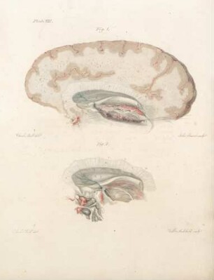
Plate VIII
Plate X
Plate I
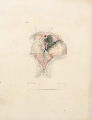
Plate VI
Plate IX
Plate III
Plate V
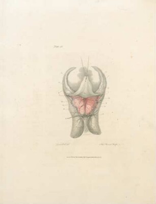
Plate IV
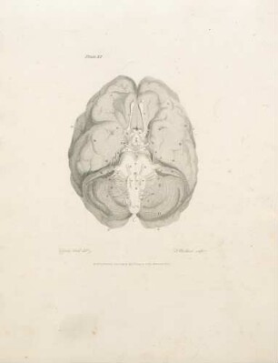
Plate XI
Plate VII
Plate II

21: The quarterly journal of pure and applied mathematics
Plate VIII
Plate X
Plate I
Plate VI
Plate IX
Plate III
Plate V
Plate IV
Plate XI
Plate VII
Plate II
21: The quarterly journal of pure and applied mathematics
Plate VIII
Plate X
Plate I
Plate VI
Plate IX
Plate III
Plate V
Plate IV
Plate XI
Plate VII
Plate II