Grafik
Plate VII
This Plate represents the simple section of the Brain, and Bones of the Face, and from it much of the relation of the parts and their general connection may be understood. The Scull is cut a little to the left of the course of the longitudinal Sinus, and the incision of the Brain is continued so as to lay open the lateral Ventricles without injuring the Septum Lucidum, or Fornix; to expose the third Ventricle also, and to give a section of the Pons Varolii, and Arbor Vitas; in short, to make a full section of the Cerebrum and Cerebellum. a. a. The cut edge of the Cranium. b. The Frontal Sinus. c. The Cethmoid Cells. d. The Antrum Highmorianum. e. The cuneiform process of the Occipital bone, where it goes forward to join the Sphenoid bone. f. The internal medullary part of the Cerebrum, or, as seen in the former sections, the Centrum Ovale of Vieussens. g. The Cineritious or Cortical Substance of the Cerebrum, into which the Pia Mater, and some of the turns of the injected Arteries, are seen to penetrate. h. The Corpus Callosum, sometimes called the Commissura Magna—we have to observe its striated or rather fibrous appeaance, and we understand the manner in which it covers the lateral Ventricles, while there descends from the middle part of it the Septum Lucidum dividing those Ventricles. i. That part of the lateral Ventricle which lies above the Fornix, being the shaded part, while the letter i stands directly upon the partition betwixt the left and right lateral Ventricles, viz. the Septum Lucidum. k. A vein stretching from the most anterior part of the Velum Interpositum, and from under the anterior Crus of the Fornix, to the fore part of the lateral Ventricle. I. The Fornix, and in this view we shall understand how this medullary body forms a floor to this upper part of the lateral Ventricle, while it stretches over the third Ventricle. m. The Posterior Crus of the Fornix of the left side cut off where it is about to turn down into the inferior part of the lateral Ventricle. {Plate III. k. and c.) n. The Left Anterior Crus of the Fornix. o. The Right Anterior Crus of the Fornix. p. The Anterior Commissure of the Brain. It seems high compared with the Crus of the Fornix, from the latter falling down. Q. The Velum Interpositum stretching under the Fornix, and covering the Thalami Nervorum Opticorum. r. The Third Ventricle, it being observed that the letter is opposed to the convex surface of the Thalami Nervi Optici of the right side, and it cannot be misunderstood that it is the cavity betwixt the surface of this and the left Thalamus (which is taken away) which forms the third Ventricle; while upon the upper part of this body, as it now appears to us, the two surfaces are united by the Commissura Mollis leaving an opening on each extremity of this adhesion, viz. Vulva and Anus, under the anterior of which is the beginning of the Infundibulum, and under the posterior the Iter ad quartum Ventriculum. s. The beginning of the Infundibulum. t. Iter ad quartum Ventriculum. x. The communication betwixt the lateral Ventricle of the right side, and the third Ventricle. z. The Pineal Gland, lying enveloped in the Velum, and inclining backwards. 1. The Commissura Cerebri posterior, which to me appears as the reflected medullary substance of the Nates, and not in any degree resembling in the section the anterior one, nor a nervous cord. The little peduncle connecting the Pineal Gland will be observed, and the little transverse medullary cords upon its base. 2. The proper Pedunculi of the Pineal Gland, which pass round upon the convex surface of the Thalami Nervorum Opticorum, and join the anterior pillar of the Fornix. These two Pedunculi are best seen after separating the Velum, and looking down upon the Optic Thalami. 3. The Tentorium, which is seen to stretch over the Cerebellum, and to support the posterior lobes of the Brain. 4. The Pia Mater continued in betwixt the Cerebrum and Cerebellum, and which connects the Nates and Testes to the Cerebellum. 5. The Nates, 6. The Testes, "We have to observe, that these eminences are not within the cavities of the Brain ; but that they may be seen by separating the Brain and Cerebellum from behind. 7. The origin of the fourth Nerve of the Brain, or Trochlears. 8. Section of the Tuber Annulare, or Pons Varolii, where the appearance of the striae, or filaments, is accurately represented. 9. The Crura Cerebelli, and Arbor Vitje; the Crura Cerebelli being formed by the union of the branches of the internal medullary part of the Cerebellum, which branching is called the Arbor Vitae. 10. The Medulla Oblongata, being the upper part of the spinal marrow, as formed by the union of the Cerebrum and Cerebellum, and enumerated commonly as one of the three great divisions of the Brain. * The Inferior Lobulus of the Cerebellum. 11. The Basilar Artery, which is formed by the union of the Vertebral Arteries. 12. The Internal Carotid Artery, where it is passing through its foramen in the sphenoid bone. 13. The Opthalmic Artery, derived from the Internal Carotid Artery within the Scull.
- Standort
-
Universitätsbibliothek Heidelberg
- Sammlung
-
UB Anatomische Illustrationen
- Inventarnummer
-
P 1242-4 Folio RES
- Sprache
-
Englisch
- Klassifikation
-
Stich (Gattung)
- Bezug (was)
-
Anatomie
Gehirn
Illustration
- Ereignis
-
Herstellung
- (wo)
-
London
- (wann)
-
um 1802
- Ereignis
-
Veröffentlichung
- (wer)
-
Longman
- (wann)
-
1802
- Ereignis
-
Veröffentlichung
- (wo)
-
London
- Letzte Aktualisierung
-
26.03.2025, 09:43 MEZ
Datenpartner
Ruprecht-Karls-Universität Heidelberg. Universitätsbibliothek. Bei Fragen zum Objekt wenden Sie sich bitte an den Datenpartner.
Objekttyp
- Grafik
Beteiligte
Entstanden
- um 1802
- 1802
Ähnliche Objekte (12)
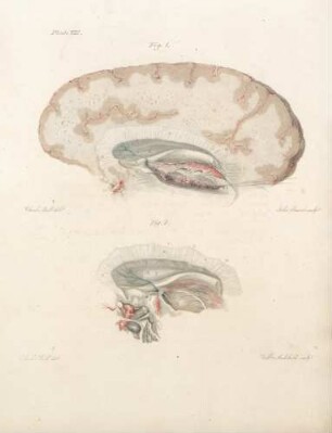
Plate VIII
Plate X
Plate I
Plate VI
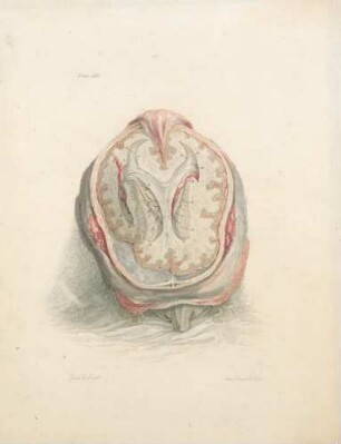
Plate III
Plate IX
Plate V
Plate IV
Plate XI
Plate II
Plate XII

21: The quarterly journal of pure and applied mathematics
Plate VIII
Plate X
Plate I
Plate VI
Plate III
Plate IX
Plate V
Plate IV
Plate XI
Plate II
Plate XII
21: The quarterly journal of pure and applied mathematics
Plate VIII
Plate X
Plate I
Plate VI
Plate III
Plate IX
Plate V
Plate IV
Plate XI
Plate II
Plate XII