Grafik
Plate III
We have in this Plate a very extensive view of the Ventricles, or Cavities of the Brain ; but to follow the appearances of the parts, as they present themselves to us during dissection, we must attend, in the first place, to the left side of the Brain. For here the Corpus Callosum is cut away, and a horizontal incision is made of the cen- tral medullary part of the Cerebrum, so as to lay open the lateral Ventricles. While upon the right side it is considerably more cut away, to expose the whole extent of the lateral Ventricle. Parts seen upon first laying open the lateral ventricle: a. The Corpus Striatum of the left side, which forms a convex floor to this part of the highest level of the lateral Ventricle. It has, like the surface of the Brain, cineritious matter without, while the section of it shows medullary striae. b. The Choroid Plexus, leading anteriorly to the communication of the Ventricles under the Fornix, while it will be seen sinking backwards into the great inferior horn of the Ventricle. c. The margin of the posterior Crura of the Fornix. d. The Tenia Semicircularis, or Tenia Striata. It will he observed to be covered anteriorly by a layer of a transparent cineritious coloured substance, viz. lame cdrnee. e. The Anterior Sinus of the lateral Ventricle, being formed by the termination forwards of the Corpus Striatum. Upon completing the section backwards we find these parts. f. The Posterior Sinus of the lateral Ventricle, which is a triangular cavity, stretching in a curved direction into the posterior lobe of the Cerebrum. This part of the Ventricle varies much in different bodies, and even sometimes the right and left sides of the same subject differ in figure and direction. g. The Cornu Ammonis, or Hippocampus Major n, in which relief or convexity the posterior horn of the Ventricle is seen to terminate, while it is, at the same time, continuous with the next eminence. h. h. The Colliculus, or l'Ergot, or Hippocampus Minor, which is a convexity or elevation in the floor of this prolonged part of the Ventricle, resembling that which descends into the great inferior, but which is sometimes called the posterior, horn of the Ventricle. Its surface consists of white medullary matter; but when the knife penetrates this cortex, it is seen to have cineritious matter within. i. k. c. The Fornix, or Vault of Three Pillars. It has this name, not from its relation to the lateral Ventricles, but from the manner in which it covers the third Ventricle. Posteriorly at c. k. we see it joining with the back part of the Corpus Callosum, and expanding what are called its posterior crura, into a broad lamina of medullary matter, which connects them with the eminences g. h. l. The Sinus, or Cavity of the Septum Lucidum. The remains of the Septum Lucidum are seen to form a ridge upon the middle part of the Fornix, for it is a partition which reaches down from the Corpus Callosum to the Fornix, upon which it seems to rest, and thus divides the two lateral Cavities or Ventricles. It consists of a double medullary lamina21, and the space betwixt these is called the Cavity of the Septum Lucidum, in general containing a serous fluid. From the varying descriptions of authors it must have great diversity in size. Vieussens and Winslow describe it as communicating with the third Ventricle. Tarin says, that it sometimes opens into the lateral Ventricle. m. The Thalamus Nervi Optici of the right side. n. The Choroid Plexus of the right side dissected up from its natural seat, and part of it cut away. Its further progress downwards into the great inferior horn of the Ventricles lies within the circle of the Cornu Ammonis, and where it receives the lower and Anterior Choroid Arteries. o. The Inferior Horn of the Lateral Ventricle, which is to be seen only by cutting the middle lobe of the Cerebrum obliquely ; for this part of the Ventricle lies very deep, and almost under the anterior Sinus, E. p. The termination of the Cornu Ammonis, which is the relief con- tinued down upon the bottom of the great inferior horn, as we see the eminence h. continued into the Processus Digitalis, or posterior Sinus.
- Standort
-
Universitätsbibliothek Heidelberg
- Sammlung
-
UB Anatomische Illustrationen
- Inventarnummer
-
P 1242-4 Folio RES
- Sprache
-
Englisch
- Klassifikation
-
Stich (Gattung)
- Bezug (was)
-
Anatomie
Gehirn
Illustration
- Ereignis
-
Herstellung
- (wo)
-
London
- (wann)
-
um 1802
- Ereignis
-
Veröffentlichung
- (wer)
-
Longman
- (wann)
-
1802
- Ereignis
-
Veröffentlichung
- (wo)
-
London
- Letzte Aktualisierung
-
26.03.2025, 09:43 MEZ
Datenpartner
Ruprecht-Karls-Universität Heidelberg. Universitätsbibliothek. Bei Fragen zum Objekt wenden Sie sich bitte an den Datenpartner.
Objekttyp
- Grafik
Beteiligte
Entstanden
- um 1802
- 1802
Ähnliche Objekte (12)
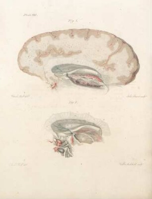
Plate VIII
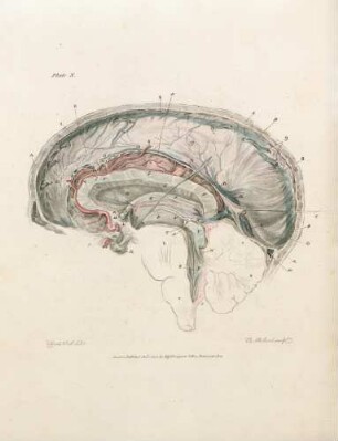
Plate X
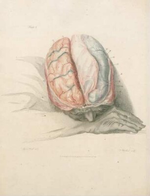
Plate I
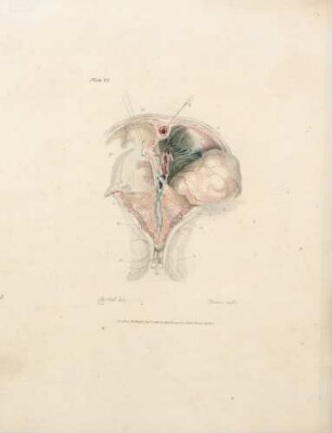
Plate VI
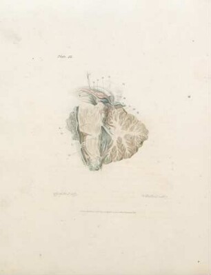
Plate IX
Plate V
Plate IV
Plate XI
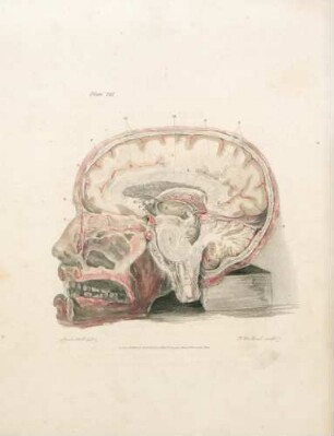
Plate VII
Plate II
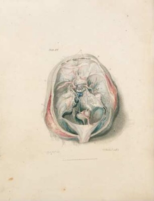
Plate XII

21: The quarterly journal of pure and applied mathematics
Plate VIII
Plate X
Plate I
Plate VI
Plate IX
Plate V
Plate IV
Plate XI
Plate VII
Plate II
Plate XII
21: The quarterly journal of pure and applied mathematics
Plate VIII
Plate X
Plate I
Plate VI
Plate IX
Plate V
Plate IV
Plate XI
Plate VII
Plate II
Plate XII