Grafik
Plate XI
This Plate explains the Base of the Brain, and is taken from Vicq. d'Azyr. GENERAL DIVISION OF THE BRAIN SEEN IN THE BASE. a. a. The Anterior Lobes of the Cerebrum. b. b. The Middle Lobes of the Cerebrum. c. c. The Posterior Lobes of the Cerebrum. d. The Cerebellum. e. The Medulla Oblongata, formed by prolongations of the Cerebrum and Cerebellum. f. The Pons Varolii, or Tuber Annulare. g. g. The Crura Cerebri white and fibrous, and formed by the Internal Medullary part of the Cerebrum, continued into the Medulla Oblongata. h. The Crura Cerebelli, prolonged in the same way from the Cerebellum into the Pons Varolii, and Medulla Oblongata, i. An Eminence which Vicq. d'Azyr calls Lobulus Medulla Oblongata. k. External and Superior Lobes of the Cerebellum. I. A Sulcus betwixt the Lobes of the Cerebellum, in which a little Falx, resembling the Falx Cerebri lies. m. Foramen Caecum Posterius. n. The Fossa Silvii, dividing the anterior and middle Lobes of the Cerebrum. 0. The Monticulus Vesalii. p. The Fossa of the Nervi Motores Oculorum, according to Vicq. d'Azyr. q. The Infundibulum. r. The Eminentije Candicantes. s. What Vicq. d'Azyr calls the " Substance perforce," which is a medullary part, perforated with many Arteries. t. The Corpora Pyramidalia. v. v. The Corpora Olivaria. 1. 1. The First Pair of Nerves, or Olfactory Nerves. 1. 2. The Second Pair of Nerves, or OpticNerves. 3. 3. The Third Pair of Nerves, or Motores Oculorum. 4. 4. The Fourth Pair of Nerves, or Trochleares. 5. 5. The Fifth Pair of Nerves, or Trjgemini. 6. 6. The Sixth Pair of Nerves, the Abducentes. 7. 7. The Seventh Pair of Nerves, consisting of two portions. The Portio Mollis, acaustic or auditory Nerve, and the Portio Dura, or Nervous Communicans Faciei. 8. 9. 8. 9. The Eighth Pair of Nerves, 8. 8. being the Fascicali from which is derived the Par Vagum, and Glosso-pharingeal Nerve. 9. 9. The Accessory Nerve of Willis. 10. 10. The Ninth Pair, or Laryngeal Nerve.
- Location
-
Universitätsbibliothek Heidelberg
- Collection
-
UB Anatomische Illustrationen
- Inventory number
-
P 1242-4 Folio RES
- Language
-
Englisch
- Classification
-
Stich (Gattung)
- Subject (what)
-
Anatomie
Gehirn
Illustration
- Subject (who)
- Event
-
Herstellung
- (who)
- (where)
-
London
- (when)
-
um 1802
- Event
-
Veröffentlichung
- (who)
-
Longman
- (when)
-
1802
- Event
-
Veröffentlichung
- (where)
-
London
- Last update
-
26.03.2025, 9:43 AM CET
Data provider
Ruprecht-Karls-Universität Heidelberg. Universitätsbibliothek. If you have any questions about the object, please contact the data provider.
Object type
- Grafik
Associated
Time of origin
- um 1802
- 1802
Other Objects (12)
Plate VIII
Plate X
Plate I
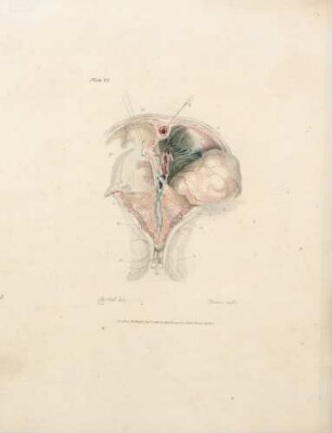
Plate VI
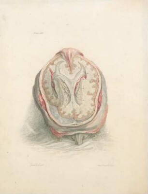
Plate III
Plate IX
Plate V
Plate IV
Plate VII
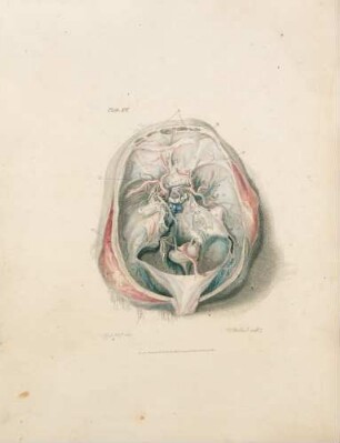
Plate XII
Plate II

21: The quarterly journal of pure and applied mathematics
Plate VIII
Plate X
Plate I
Plate VI
Plate III
Plate IX
Plate V
Plate IV
Plate VII
Plate XII
Plate II
21: The quarterly journal of pure and applied mathematics
Plate VIII
Plate X
Plate I
Plate VI
Plate III
Plate IX
Plate V
Plate IV
Plate VII
Plate XII
Plate II