Grafik
Plate IV
In this Plate the parts are nearly of their natural size. It is an enlarged view of what is seen in the third Plate within the lateral Ventricles, while the Fornix is here lifted up, showing the attachment of the Choroid Plexus, and the Velum Interpositum of Haller. When we have the parts in the situation represented in the last Plate, we shall, by following the Choroid Plexus, B, forward to where it leads under the anterior pillar of the Fornix, find the com- munication of the Ventricles. If we direct our probe horizontally, and under the anterior pillar of the Fornix, wc lind it has an easy passage into the lateral Ventricle of the other side; if downwards, we find it descending into the third Ventricle. When we have passed the probe betwixt the two lateral Ventricles, if we lift up the anterior pillars of the Fornix, as in this Plate, we find the probe lying in the anterior part of the third Ventricle, as described by some authors, but more properly in the Foramen Commune Anterius. a. The Corpora Striata. b. b. The Anterior Sinus, or horn of the lateral Ventricle. c. c. The Posterior Horn, or Processus Digitalis. d. d. The Tenia Striata, or Centrum Semicirculare Geminum of Vieussens. e. The Fornix, cut from the anterior Crura, separated from the Velum Interpositum, and held up. f. f. The Anterior Crura of the Fornix, connected with g. The Comissura Cerebri Anterior. h. The Lyra, that is simply the inferior surface of the Fornix, which in the natural situation of the parts lies upon the Velum Interpositum. i. i. The Corpora Fimbriata, which is the edge of the Medullary Lamina, and which is extended from the Posterior Pillars, or crura of the Fornix, and continued along the Circle of the Cornua Ammonis on each side. This is called also the Tenia Hippocampi. l. l. The Colliculus, or Hippocampus Minor. m. m. The Velum Interpositum, or Toile Choroidiene, which is a process of the Pia Mater, expanded betwixt the Fornix and third Ventricle, and which lies upon, and is attached to the Thalami Nervorum Opticorum. n. n. The Choroid Plexus, the connection of which with the Velum is now understood. The Choroid Plexus seems, upon the first view, before the Fornix is lifted, to be a congeries of vessels unconnected with the floor of the Ventricle; but upon unravelling and spreading it with the probe, it is found to be a membrane, and to be continuous with the Pia Mater, lining the floor of the Ventricle, and is, in fact, merely the Vascular Pia Mater gathered together into folds. And, upon raising the Fornix, we find it connected with the Velum Interpositum as with a mesentery. o. p. p. Branches of the Vena Magna Galeni, which beingdistributed to the Corpora Striata, and paries of the Ventricle, pass under the Tenia Striata, and run into the Velum Interpositum, under the Fornix. A piece of cord waxed, so as to give something of the stiffness of a probe. It is introduced into the communication betwixt the Ventricles, and upon lifting the Fornix is seen lying in the upper and fore part of the third Ventricle, or rather the Foramen Commune Anterius.
- Location
-
Universitätsbibliothek Heidelberg
- Collection
-
UB Anatomische Illustrationen
- Inventory number
-
P 1242-4 Folio RES
- Language
-
Englisch
- Classification
-
Stich (Gattung)
- Subject (what)
-
Anatomie
Gehirn
Illustration
- Subject (who)
- Event
-
Herstellung
- (who)
- (where)
-
London
- (when)
-
um 1802
- Event
-
Veröffentlichung
- (who)
-
Longman
- (when)
-
1802
- Event
-
Veröffentlichung
- (where)
-
London
- Last update
-
26.03.2025, 9:43 AM CET
Data provider
Ruprecht-Karls-Universität Heidelberg. Universitätsbibliothek. If you have any questions about the object, please contact the data provider.
Object type
- Grafik
Associated
Time of origin
- um 1802
- 1802
Other Objects (12)
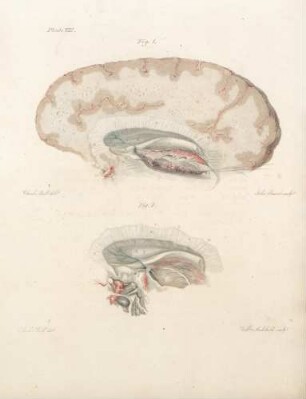
Plate VIII
Plate X
Plate I
Plate VI
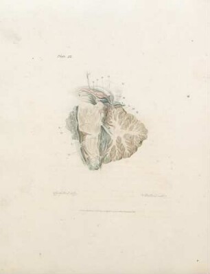
Plate IX
Plate III
Plate V
Plate XI
Plate VII
Plate II
Plate XII

21: The quarterly journal of pure and applied mathematics
Plate VIII
Plate X
Plate I
Plate VI
Plate IX
Plate III
Plate V
Plate XI
Plate VII
Plate II
Plate XII
21: The quarterly journal of pure and applied mathematics
Plate VIII
Plate X
Plate I
Plate VI
Plate IX
Plate III
Plate V
Plate XI
Plate VII
Plate II
Plate XII