zweidimensionales bewegtes Bild
Topographische Anatomie des menschlichen Embryos nach Entwicklung der ersten Nerven
Totalrekonstruktion eines 3,5 mm großen menschlichen Embryos. Am Modell werden Gehirnanlage, Hals-Rumpfregion, Herzanlage sowie die Anlage von Gallenblase und Leber im einzelnen demonstriert. Weitere Aufnahmen zeigen das Blutgefäßsystem und einige endokrine Drüsen.
Total reconstruction of a 3,5 mm human embryo. Brain anlage, neck-trunk region, heart anlage, anlage of gallbladder and liver are demonstrated using a model. Further shots show the vascular system and some endocrine glands.
- Alternative title
-
Topographical Anatomy of the Human Embryo after Development of the First Nerves
- Location
-
Hannover TIB
- Extent
-
269MB, 00:04:32:00 (unknown)
- Language
-
Deutsch
- Notes
-
Audiovisuelles Material
- Bibliographic citation
-
Topographische Anatomie des menschlichen Embryos nach Entwicklung der ersten Nerven ; (Jan. 1970)
- Keyword
-
Medizin
Nervensystem / Embryonalentwicklung
Embryonalentwicklung
embryology
embryo / anatomy
Embryologie
embryo / histology
Embryonalentwicklung / Mammalia
Gehirnanlage
brain anlage
embryonic development / Mammalia
medicine
- Event
-
Veröffentlichung
- (who)
-
IWF (Göttingen)
- (when)
-
1970-01-01
- Contributor
-
Blechschmidt, Erich
- DOI
-
10.3203/IWF/W-909
- Last update
-
04.12.2024, 8:09 AM CET
Data provider
This object is provided by:
Technische Informationsbibliothek (TIB). If you have any questions about the object, please contact the data provider.
Technische Informationsbibliothek (TIB). If you have any questions about the object, please contact the data provider.
Object type
- zweidimensionales bewegtes Bild
Associated
- Blechschmidt, Erich
- IWF (Göttingen)
Time of origin
- 1970-01-01
Other Objects (12)
Bewegungsspiel des Rückens - Anatomie des Lebenden

Tonschwingungen am überlebenden menschlichen Mittelohrapparat
Die weiße Substanz des menschlichen Gehirns
Die basalen Ganglien des menschlichen Gehirns
Häute und Oberfläche des menschlichen Gehirns
Die grüne Stadt oder "Vom menschlichen Maß"
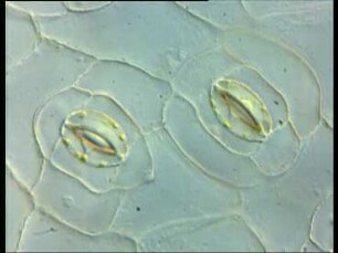
Spaltöffnungen und Regulation der Photosyntheseaktivität
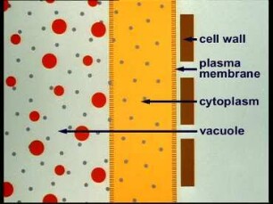
Plasmolysis and Deplasmolysis
Nordeuropa, Ostnorwegen - Spielen auf der Hardanger-Geige

Des Lebens Wunderhorn
Stomata and regulation of photosynthetic activity
Nordeuropa, Südnorwegen - Herstellen von Silberfiligran
Bewegungsspiel des Rückens - Anatomie des Lebenden
Tonschwingungen am überlebenden menschlichen Mittelohrapparat
Die weiße Substanz des menschlichen Gehirns
Die basalen Ganglien des menschlichen Gehirns
Häute und Oberfläche des menschlichen Gehirns
Die grüne Stadt oder "Vom menschlichen Maß"
Spaltöffnungen und Regulation der Photosyntheseaktivität
Plasmolysis and Deplasmolysis
Nordeuropa, Ostnorwegen - Spielen auf der Hardanger-Geige
Des Lebens Wunderhorn
Stomata and regulation of photosynthetic activity