zweidimensionales bewegtes Bild
Gutartige und bösartige Pigmentzellen
Anatomische und histologische Grundlagen. Entstehung eines Melanoms aus einem Nävus-Zellnävus scheinbar gesunder Haut. Differentialdiagnostische Abgrenzung der Melanome: Naevus pigmentosus, Keratosis senilis, Verruca seborrhoica, pigmentiertes Basaliom, Naevus coerulens, Angiome und als Melanoma in situ der Morbus Dubreuilh. Klinische Bilder des Melanoms: in der Tiefe der Haut liegende, oberflächlich flache oder exophytisch wachsende Tumoren. Nah- und Fernmetastasen des Primärtumors (Trick, Röntgenaufnahmen, histologische Schnittbilder, Gewebekulturen).
In human skin melanocytes and nevus cells are differentiated. Melanocytes may undergo malignant change and may become melanoma cells. Their high mobility is made visible by microcinematography. The first part of the film concerns itself with manifestations on the skin which are due to usually benign nevus cells. Alterations of the skin which must be regarded as melanomas are shown in the second part. Finally, the author refers to treatment of melanoma. Subsequent to the therapeutic part proximal and distal metastases of the melanoma are shown.
- Weitere Titel
-
Benign and Malignant Pigment Cells
- Standort
-
Hannover TIB
- Umfang
-
203MB, 00:16:31:11 (unknown)
- Sprache
-
Deutsch
- Anmerkungen
-
Audiovisuelles Material
- Erschienen in
-
Gutartige und bösartige Pigmentzellen ; (Jan. 1970)
- Schlagwort
-
skin cancer
nevus cells
Medizin
Hautkrebs
Melanom
Melanozyten
melanoma / diagnosis
melanoma / secondary
Nävuszellen
skin neoplasms
melanocytes
medicine
- Ereignis
-
Veröffentlichung
- (wer)
-
IWF (Göttingen)
- (wann)
-
1970-01-01
- Beteiligte Personen und Organisationen
-
Rohde, Bernward
- DOI
-
10.3203/IWF/C-1022
- Letzte Aktualisierung
-
04.12.2024, 08:09 MEZ
Datenpartner
Technische Informationsbibliothek (TIB). Bei Fragen zum Objekt wenden Sie sich bitte an den Datenpartner.
Objekttyp
- zweidimensionales bewegtes Bild
Beteiligte
- Rohde, Bernward
- IWF (Göttingen)
Entstanden
- 1970-01-01
Ähnliche Objekte (12)
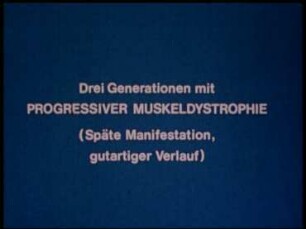
Drei Generationen mit progressiver Muskeldystrophie (späte Manifestation, gutartiger Verlauf)

Differentialdiagnose gutartiger und bösartiger Dickdarmerkrankungen
Spaltöffnungen und Regulation der Photosyntheseaktivität
Stomata and regulation of photosynthetic activity
Des Lebens Wunderhorn
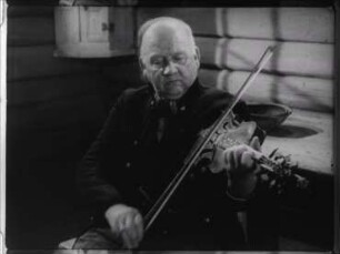
Nordeuropa, Ostnorwegen - Spielen auf der Hardanger-Geige
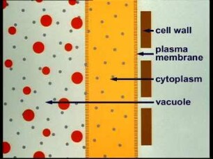
Plasmolysis and Deplasmolysis
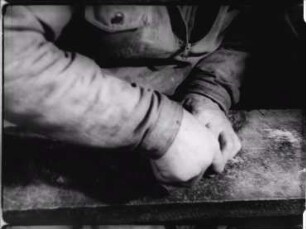
Nordeuropa, Ostnorwegen - Herstellen von Messern
Nordeuropa, Südnorwegen - Herstellen von Silberfiligran
Bösartige Geschwülste in histologisch gutartigem Habitus
Gutartige und bösartige Weichteiltumoren der zentralen Körperhöhlen
Differentialdiagnose der gutartigen und bösartigen Erkrankungen der Ileozökalgegend
Drei Generationen mit progressiver Muskeldystrophie (späte Manifestation, gutartiger Verlauf)
Differentialdiagnose gutartiger und bösartiger Dickdarmerkrankungen
Spaltöffnungen und Regulation der Photosyntheseaktivität
Stomata and regulation of photosynthetic activity
Des Lebens Wunderhorn
Nordeuropa, Ostnorwegen - Spielen auf der Hardanger-Geige
Plasmolysis and Deplasmolysis
Nordeuropa, Ostnorwegen - Herstellen von Messern
Nordeuropa, Südnorwegen - Herstellen von Silberfiligran
Bösartige Geschwülste in histologisch gutartigem Habitus
Gutartige und bösartige Weichteiltumoren der zentralen Körperhöhlen